
The talus heals slowly. It takes two to three months before the patient is allowed to walk and put weight on the talus again, after the fracture. Once the talus bone heals, the function of the ankle should be boosted through exercise and physical therapy.
Full Answer
How to engage the talus and calcaneus posteriorly?
Aug 01, 2007 · Treatment. The goals of treatment are to restore the normal anatomic relationships between the talus, the navicular, and the calcaneus, in order to provide a normal weight distribution through the foot. Treatment of congenital vertical talus, as with clubfoot, begins with serial manipulations and casts.
What are the goals of treatment for congenital vertical talus?
Feb 04, 2021 · Stretching exercises within a physical therapy/rehabilitation program will help you restore the range of motion, strength, and stability of your calcaneus after the bones have adequately healed. Complications Complications can often occur with calcaneus fractures both when the fracture happens and during and after surgery.
What is the problem with the talus bone?
Talus or Talocrural Joint Mobilization Key Concept: You must draw the Inferior Calcaneus Anteriorly when you are mobilizing the Talus posteriorly. This will engage the Talus and Calcaneus and will enhance the rotational aspect of Talar mobility. You cannot just glide the Talus posteriorly, the Talus must also rotate with the Calcaneus.
What is the treatment for a talus fracture?
Non-operative treatment generally involves immobilization and no weight bearing for 6 to 12 weeks followed by progressive weight bearing and ROM exercises. Operative treatment generally involves open reduction and internal fixation (ORIF) followed by immobilization, no weight bearing and ROM exercises.
How do I strengthen my ankle talus?
2:565:20Improve Your Ankle Mobility INSTANTLY with This Simple ...YouTubeStart of suggested clipEnd of suggested clipAnd further into dorsiflexion I'd do three sets of ten on the affected side of course if both anklesMoreAnd further into dorsiflexion I'd do three sets of ten on the affected side of course if both ankles are tied you could do three sets of ten on each side.
How do you strengthen a subtalar joint?
Exercise to improve subtalar joint pronation. When it can't go any further, start to bring the outside of your heel back up towards the outside of your shin. Hold the top position for a second or two before slowly lowering your foot back down towards the floor.Jul 15, 2020
How do you self mobilize the talus?
3:014:47Self Ankle Mobilization | Week 35 | Movement Fix Monday - YouTubeYouTubeStart of suggested clipEnd of suggested clipBecause those are the things working here and then I'll go again pull pull pull pull pull. So that'sMoreBecause those are the things working here and then I'll go again pull pull pull pull pull. So that's a way to then use some of the musculature in the lower leg to create ankle dorsiflexion.
How do you rehab an unstable ankle?
1:3110:49Chronic Ankle Instability Rehab and Ankle Sprain Therapy - YouTubeYouTubeStart of suggested clipEnd of suggested clipIf you're out on the trail. And what we want to do is lock that ankle into position and now try toMoreIf you're out on the trail. And what we want to do is lock that ankle into position and now try to move that ankle out of that position.
How do you crack a subtalar joint?
0:552:07Exercise strategies for the subtalar joint - YouTubeYouTubeStart of suggested clipEnd of suggested clipI can also have to take a step forward but those pack in just wring your hands to the right toMoreI can also have to take a step forward but those pack in just wring your hands to the right to charge. Again. Again you can see his left foots going through a lot of really.
How do you release a subtalar joint?
0:321:58Subtalar Joint Traction | Assessment & Mobilization - YouTubeYouTubeStart of suggested clipEnd of suggested clipAnd middle finger embrace the neck of the tailless. From ventral and squeeze your patients calcaneusMoreAnd middle finger embrace the neck of the tailless. From ventral and squeeze your patients calcaneus with the FINA eminence of both of your hands you can fixate this position by crossing your thumbs.
How do you train for ankle mobility?
Ankle jumpsStand straight with your hands on your hips.Jump up straight without bending your knees.Flex your ankles, and pull up your toes while you're in the jump (dorsiflex).Extend your ankles back just before you touch the floor.Push the balls of your feet into the floor explosively, and then jump again.More items...•May 28, 2019
What is dorsiflexion of the ankle?
Dorsiflexion is the backward bending and contracting of your hand or foot. This is the extension of your foot at the ankle and your hand at the wrist. You can also dorsiflex your fingers and toes, though usually the term is referring to your wrist or ankle.
How do I get more ankle dorsiflexion?
Place the foot of the leg that is not kneeling and place it about five inches away from the wall. Lean into that front leg. Without moving your foot, try to get your knee to touch the wall. If it touches, your dorsiflexion is not too bad.
How do I strengthen my calcaneus?
4. Wall calf stretchStand a few feet from a wall with your left foot in front of your right foot.Lean toward the wall as you bend your left knee slightly.Slowly place your weight into your left foot.Keep your right knee straight as you lift your right heel off the ground. ... Hold this position for 15 to 30 seconds.More items...•Apr 19, 2019
How long does it take to improve ankle mobility?
How long will it take to improve your ankle mobility? Do the above exercises every day and you should feel results pretty damn quick (think 2 to 4 weeks).Jan 15, 2021
Can I fix chronic ankle instability?
Non-surgical treatments are very effective for many patients with chronic ankle instability and typically include anti-inflammatory medication, wearing a brace and seeing a physical therapist. If surgery is necessary, your surgeon may repair or tighten the ligaments that have been stretched.Sep 29, 2021
What bone is driven up against the talus?
For example: If you land on your feet from a fall, all of your body's weight is directed downward, and this drives the talus bone directly into the calcaneus. If you are involved in a motor vehicle accident, and your heel is crushed against the floorboard, the calcaneus is driven up against the talus.
How to heal an unstable calcaneus fracture?
Unstable calcaneus fractures require surgery in order to properly heal. The procedure will reposition the bone fragments into their accurate alignment (this is called reduction) and then held together in place with screws or metal plates attached to the outer part of the bone.
What are the bones of the foot called?
The foot is divided into three parts: the hindfoot, the midfoot and the forefoot. The hindfoot and midfoot are made up of seven bones called tarsals. The calcaneus (also called the heel bone) is the largest tarsal bone of the foot.
What is the most common fracture of the tarsal bone?
Calcaneus fractures are rare, although they are the most commonly fractured tarsal bone. These fractures can occur in many different situations, but most commonly occur in high-energy trauma situations such as car crashes or falls from heights (ex. a ladder or roof). Symptoms generally depend on the severity of the fracture ...
How to tell if a calcaneus fracture is cracked?
Symptoms of calcaneus fracture can include: Significant ankle pain: Especially when the heel is squeezed. Swollen an kle.
What are the symptoms of a calcaneus fracture?
Symptoms generally depend on the severity of the fracture but usually include significant pain, swelling, bruising, numbness, limited mobility, and limping, among others. Calcaneus fractures can lead to long-term deformity and require good follow-up and management.
How long does it take for a calcaneus fracture to heal?
Depending on the extent and severity of your fracture and the stability of the calcaneus, recovery can be a very long process that can last months to years. It may take even longer for the involved ligaments and tendons to heal. To further assist in your recovery, your physician may also suggest physical therapy.
What causes a fracture of the talus and calcaneus?
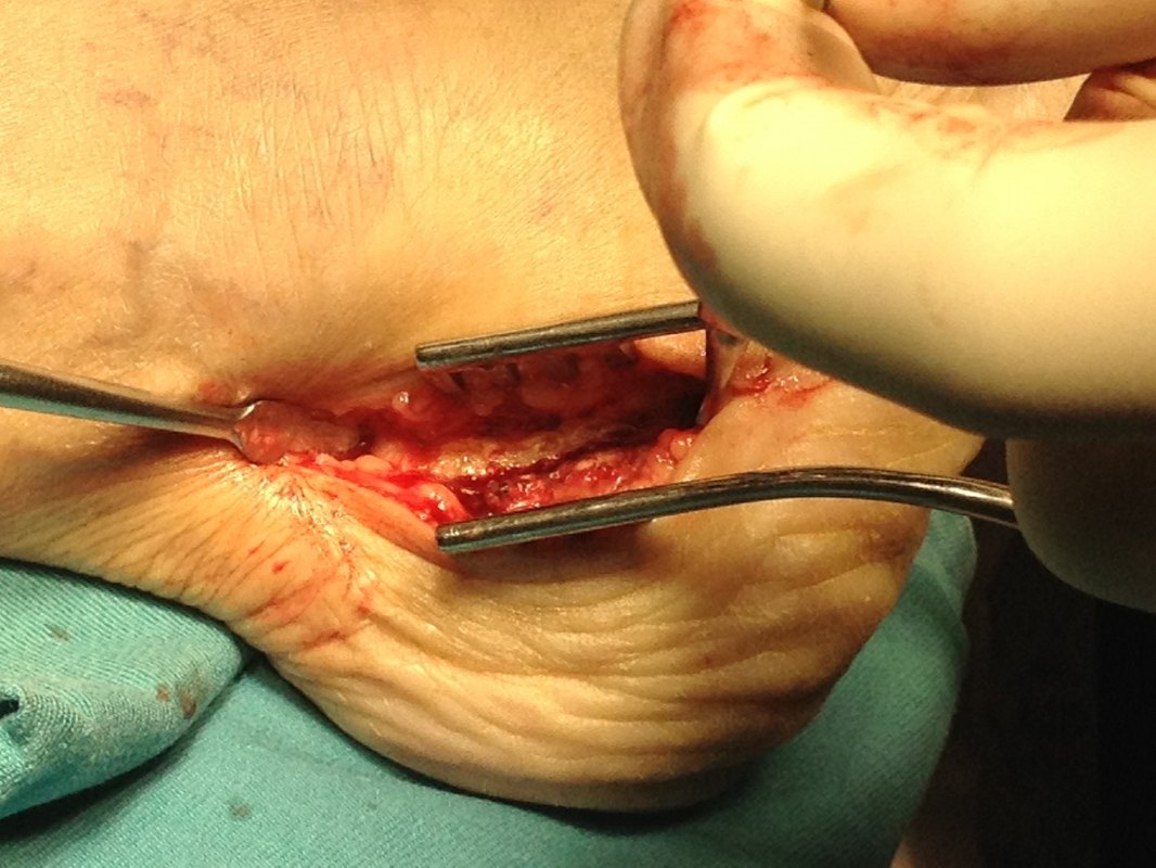
Fractures of the calcaneus and talus, collectively termed “hindfoot fractures” are typically caused by high-impact forces like falls or motor vehicle accidents. Calcaneus fractures are the more common; talus fractures, though less common are often associated with greater morbidity: owing to the bone’s tenuous blood supply of the talus, fractures there have difficulty healing. Hindfoot fractures are caused by axial load, and therefore can be seen with more proximal injuries, such as fracture of the pelvis or spine, as well.
What is the articulation between the talus and calcaneus called?
The articulation between the talus and calcaneus is called the subtalar joint. The talus does not sit on top of the center of the calcaneus, ...
What is talar neck fracture?
Talar neck fractures, caused by excessive dorsiflexion of the foot against the distal tibia, comprise half of all talus fractures. They are classified, with increasing severity, as nondisplaced; displaced but with an intact ankle joint; and displaced subluxation/dislocation of both the subtalar joint and ankles joints.
Why do people get hindfoot fractures?
Because most hindfoot fractures occur in the setting of acute injury, such as falls or motor vehicle accidents, prevention mostly centers on avoiding such accidents. However, certain health conditions can also predispose people to hindfoot fractures. For example, Kathol et al (PMID: 1871285) and Cheng et al (PMID: 9200007) found that diabetes mellitus and low bone mineral density are major risk factors for hindfoot fractures.
What is the most common injury to a calcaneal fracture?
A high-energy axial load can also cause injuries outside the lower limb. One of the most common injuries is thoracolumbar spine fractures, occurring in 10% of patients with calcaneal fractures.
What is the function of the hindfoot?
The hindfoot functions to bear and distribute weight to the foot while standing, and to permit complex foot movements in coordination with the ankle joint , especially inversion/eversion and axial rotation. The talus has a complex architecture, enabling it to function as a "ball-joint" between the leg and the foot.
Can a hindfoot fracture be missed?
Hindfoot fractures can be missed in patients who have sustained polytraumatic injuries. Thus an axial load mechanism should be a “red flag” suggesting the presence of a hindfoot fracture, and the presence of such a fracture in one limb should prompt close evaluation of the contralateral side (as it may have been subjected to the same axial load).
How to treat talus fracture?
These approaches to treatment include immobilization (making sure you can't move the bone as it heals), as by placing the foot in a cast or a boot, or surgery. Surgery, if performed, uses incisions to approach the bone which is then reduced and put back together by means of pins and screws.
How long does it take for a talus fracture to heal?
The talus heals slowly. It takes two to three months before the patient is allowed to walk and put weight on the talus again, after the fracture. Once the talus bone heals, the function of the ankle should be boosted through exercise and physical therapy. If there is loss of blood flow to the talus and avascular necrosis (dying out of the bone due to lack of blood supply) occurs, it is common to attempt surgery aimed at improvement of blood circulation to the bone.
What is the treatment for a broken ankle?
All talus fracture treatments aim to allow for maximization of the movement of the ankle and subtalar joints. Also, the goal is to restore the normal size and shape of the bone and to prevent occurrence of arthritis in both ankle and the subtalar joint later in life.#N#Planning the right treatment for a broken ankle bone can be complicated in cases of serious fractures due to their magnitude. Common complications with serious injuries include the development of arthritis or loss of blood supply to the bone. These complications can be serious, as the bone needs constant and uninterrupted blood supply in order to function properly, or in this case, to heal and regenerate after a fracture. Lack of blood supply commonly leads to arthritis or collapse of a part of the bone. A limp and chronic pain are among the complications of a talus fracture as well.
What is the talus?
The talus is directly responsible for the upward and downward movement in the ankle joint. As it joins the heel bone (the calcaneus) to form the subtalar joint, the talus is also responsible for inward and outward movement of the foot. In this way, most of your foot and ankle related motions depend on the talus.
How do your feet and legs move?
Your leg and your foot are connected by the talus. You know how your feet can move in so many directions? This is because the talus and the joint that surround the talus (the ankle joint) allow for great mobility. The talus is directly responsible for the upward and downward movement in the ankle joint. As it joins the heel bone (the calcaneus) ...
Does talus affect ankle mobility?
In this way, most of your foot and ankle related motions depend on the talus. It won't come as a surprise that any injury to the talus or ankle bone will affect your mobility to a lesser or greater extent. Injuries to the talus can be minor. For example, chips and small fragments may break off from the edge of the talus.
Can a talus fracture be minor?
Injuries to the talus can be minor. For example, chips and small fragments may break off from the edge of the talus. There are also major talus-related fractures that can be debilitating, as these effect the multiple planes of movement of the foot and ankle.
Which hand stabilizes the talus?
The Therapists aligns shoulder and arm parallel to the bottom of the foot, stabilizes the talus with the proximal hand and places the base of the distal hand on the side of the calcaneus medially to cause a lateral glide and laterally to cause. a medial glide.
Which hand grasps around the calcaneus from the pos terior aspect of the foot?
The distal hand grasps around the calcaneus from the pos terior aspect of the foot. The other hand fixes the talus and malleoli against the table and the calcaneus is pulled distally with respect to the long axis of the leg. Subtalar Medial Glide or Lateral Glide.
Where is the proximal hand placed on the dorsum of the foot?
To mobilize the tarsal joints along the medial aspect of the foot, Therapist positions himself on the lateral side of the foot and places the proximal hand on the dorsum of the foot with the fingers pointing medially so the index finger can be wrapped around and placed under the bone to be stabilized.
What is joint mobilization?
Joint mobilization refers to manual therapy techniques that are used to modulate pain and treat joint dysfunctions that limit range of motion (ROM) by specifically addressing the altered mechanics of the joint. The altered joint mechanics may be due to pain and muscle guarding, joint effusion,contractures or adhesions in the joint capsules or supporting ligaments, or malalignment or subluxation of the bonysurfaces.